
The evenings are getting lighter again, spring is almost within reach, we’re starting to think about coming out of hibernation … but it’s not quite warming up yet. Never fear! Here is the next instalment of “The decade in science so far…”
This month we will be exploring self driving cars, tiny robots that could deliver life saving drugs and the search for the first humans who will try to live on Mars.
Just like last month I have split the article up into sections to hopefully make it a little easier to read. Just like the February, this article is a little shorter than the previous one – turns out science slowed down a little after its hot start!
Lets start by jetting off to another planet!
Space
Setting up home on Mars
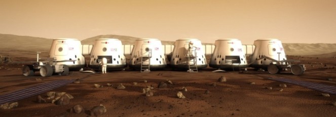
With the potential to secure a one-way ticket to Mars over 202,000 wannabe astronauts filled out the online application as part of the world wide Mars One mission.
This mission is the creation of a Dutch entrepreneur Bas Lansdorp and scientist Arno Wielders back in 2011. Applications opened in 2013 and the reason this story is featuring in this months issue is because in February 2015 Mars One announced it had a short-list of 100 of the best applicants. 50 men and 50 women from all across the world have been deemed to show the most promise of being suitable inhabitants of Mars when the first mission is launched in 2026.
The Mars One mission road map claims that they will first send unmanned missions to Mars with some proof of concept technologies to ensure it would be safe to send humans. The road map also claims that 2016 will see the start of crew training which will continue until the launch in 2026, also acting as a further selection process to ensure the first people on Mars are the best possible crew.
All sounds pretty organised right? Maybe wrong! There are a few members of the final 100 who have expressed doubt over the feasibility of the mission, prompting the article Is Mars One A Scam? Some claim they have never met anyone from the Mars one mission team, the only contact they have had being a 10 minute Skype chat.
Will people really be on a one way mission to Mars in 2026? Watch this space!
Medicine
Release the robots! Into your bloodstream!
Video Source: “Wirelessly powered, self propelled medical device” Uploaded by Stanford University School of Engineering on YouTube. Animation by Carlos Suarez, StrongBox3d
Robots are the perfect example of science fiction becoming science fact. They are becoming ever more present in our lives. I, for one, am incredibly excited by the prospect of having a little BB8 follow me around.
With all the advances in technology it is very possible that the future could include robots that are put into our bodies to deliver drugs and even perform surgeries.
The breakthrough came in February 2012 when Stanford University revealed tiny nanobots that are capable of propelling themselves through fluid, including blood. The device was created by Electrical engineer Professor Ada Poon and is powered wirelessly outside of the body. It is hoped that these devices could be used to deliver drugs, take biopsies and even perform tiny surgeries inside the body.
The plausibility of these devices comes from the ability to power and control them from outside the body. Most other devices of this kind are battery powered which makes them larger and heavier. The new nanobots work through the use of a radio transmitter outside the body that sends signals to the device.
Electricity is essential just the flow of electrons through a circuit. An electric current can be generated by passing a wire through a magnetic field. A material that is capable of conducting electricity will contain many free electrons (negatively charged sub-atomic particles), and the motion of passing the wire through a magnetic field causes the free electrons in the wire to be deflected. This creates a positive and a negative charge at different ends and because electrons are negatively charged they will move towards the positive end. The electrons start to flow and hey-presto you have a current.
The new medical devices Prof. Poon has created are magnetically coupled to a transmitter outside the body, meaning that any changes in the transmitter will induce changes in current in the device and create power. This nifty system allows it to be propelled through the blood stream.
The devices still need further development and it will be a while before they are actually used in patients, but we are definitely on our way to releasing robots into the blood stream.
(If you want to read some more about the different types of devices Prof. Poon created this article on Gizmag gives a great explanation.)
Biology
Lab grown brain that has a short term memory
Memory is a funny thing that we still don’t fully understand, playing havoc at times when we need to remember something that happened a long time ago (I wrote about this in a previous blog post: Your memory is failing you). How memory works and just generally how our brains work is a big deal in the world of scientific research, the two largest biology research councils (BBSRC and MRC) in the UK spend roughly £70 million a year on neuroscience research.
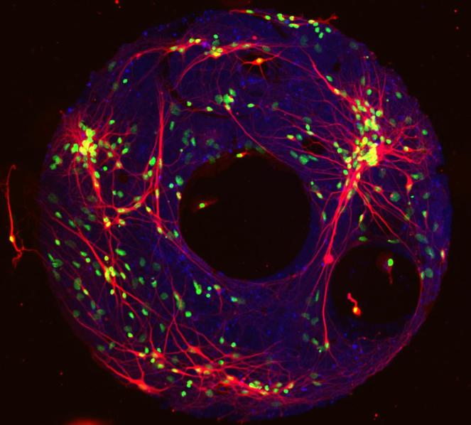
A pretty cool breakthrough was released in February 2011: tiny brain models had been created using living brain cells. The models consisted of only 40-60 neurons, a long way off the 100 billion in the average human brain, but they were still sending electrical signals around the model like neurons would in any living brain!
Created from brain cells of rat embryos by a team at the University of Pittsburgh, it was hoped the artificial ‘microbrains’ could help scientists study the electrical signals the brain sends and the way it stores information.
Not only were these cells able to connect with each other and form a network on the protein coated dish they were grown on, they also appeared to have some sort of memory function. Not to the level that we might initially think of as memory, but when stimulated with an electrical impulse the cells maintained the signal for 12 seconds. The microbrain is essentially storing the signal long (in science 12 seconds is a long time!) after the stimulus has finished.
These cells were ‘remembering’ the signal!
Original research: Ring-shaped neuronal networks: a platform to study persistent activity
The discovery of a King
Before we get to the science in this story, lets start with a quick history lesson…
Richard III was King of England from 1483 – 1485. His death marked the end of the Middle Ages. He was also the last King to be killed in battle on home soil. His remains were thought to have been buried in Greyfriars Friary Church in Leicester, however there were also reports that his remains had been discarded in a river. After the friary’s demolition in the mid 1500’s Richards remains were thought to be lost for almost 5 centuries.
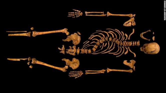
What’s all this got to do with science? When the hunt for his remains began and a human skeleton was discovered in the site of the old friary, now a car park, the race was on to try and identify who the remains could belong too. This involved some interesting science.
The first technique the team used was Carbon Dating, use to estimate the age of dead plant, animal and even human remains. A tiny proportion of the carbon in the atmosphere (one part per trillion) is radioactive and known as Carbon-14 (14C). When plants take in carbon dioxide from the atmosphere some of this is 14C, which is passed through the food chain into humans. When we die the exchange of carbon to the environment stops and gradually the amount in the body decreases due to radioactive decay. 14C has been well studied and we know how long it takes for it to loose half of its radioactivity, known as its half life. For 14C its 5,730 years. The team trying to identify King Richard were therefore able to date the bones by measuring how much 14C was left. Two of the carbon dating results obtained from the remains dated to 1430-1460 and 1412-1449, too old to be Richard. However there are many factors that can effect the estimations of carbon dating, including diet. Analysis of teeth showed that the person had a rich seafood diet, taking this into account it was predicted with 95% certainty that the remains were from 1450-1540, the right time for these remains to be Richard.
Along with carbon dating genetic tests were also carried out to try and determine the identity of the remains. To do this they had to obtain mitochondrial DNA (mtDNA) from the remains. Mitochondria are an organelle found in cells that are responsible for energy turn over, through the process of respiration, and the mtDNA is passed directly from mother to child, so can be traced through the maternal line. Luckily the family tree of nobel families are very well kept and they were able to compare the mtDNA with that of a living maternal line relative of King Richard III. It was a match!
This DNA evidence, along with many characteristics of the skeleton matching historical records of Richard’s appearance, lead the team at the University of Leicester to announce in February 2013 that they had found the remains of the late King. Not everyone was convinced however; there was doubt cast over the emphasis given to the mtDNA match. This could have been any descendent of Richard’s mother or either of her sisters and the carbon dating only gives very broad era. There was also scepticism due to the lack of peer reviewed evidence in scientific journals at the time of the announcement.
The University of Leicester did eventually publish the genetic finding in Nature Communications in December 2014. Personally I think it’s quite a convincing read.
I guess we will never know for sure if Richard III did lay under a car park in Leicester for over 500 years, but it seems pretty likely.
Physics
Live imaging the brain
The majority of what scientists have learned about the functional workings of the brain comes from looking at dead cells and trying to piece together what would be happening in the living cells, or by growing brain cells in culture which can’t be guaranteed to act exactly how they would in the living brain.
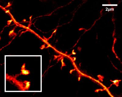
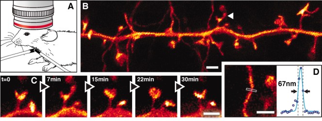
But in February 2012 that was set to change when scientists at the Max Plank Institute in Germany reported their new technique for imaging neurones in a living mouse brain. The microscope they were using is called a STED (stimulated emission depletion) microscope. This is a type of super-high resolution microscopy that works by essentially cancelling out light diffraction.
Diffraction occurs when light hits the edge of an object – the light wave bends around it and its path is distorted slightly. The amount the light that is diffracted depends of the size of the light wave and the size of the gap it is trying to go through. If the gap is bigger than the wave of light there will be no diffraction. This phenomenon can be seen in everyday life; the tracks on a CD are closely spaced and when light hits the edge of the little grooves it is diffracted, causing the rainbow appearance you can see when you hold it up to light.
This diffraction can be an issue when trying to obtain super resolution images, but STED microscopy seems to have gotten around it! It relies on the use of fluorophores, molecules that fluoresce different colours when excited by certain types of light. They are used all the time in scientific research and animal models such as mice can be genetically modified to express these molecules within certain tissues. The STED microscope uses a laser that de-excites the fluorophores in a ring shape around the spot you are trying to focus on, making the amount of light being looked at through the microscope very small – a smaller wave of light through a bigger gap = less diffraction. This technique allows the generation of super resolution images without the problem of distortion through light diffraction.
This is all well and good but there is still the fact that these microscopes are used to look at a living mouse brain! They used the genetically modified mice that I mentioned earlier, put them under anaesthetic and remove a part of the mouse skull. This is then replaced with a glass window, enabling them to see the live glowing neurones in super resolution.
Below are some of the images they snapped!
Technology
The smart robots are out to play!
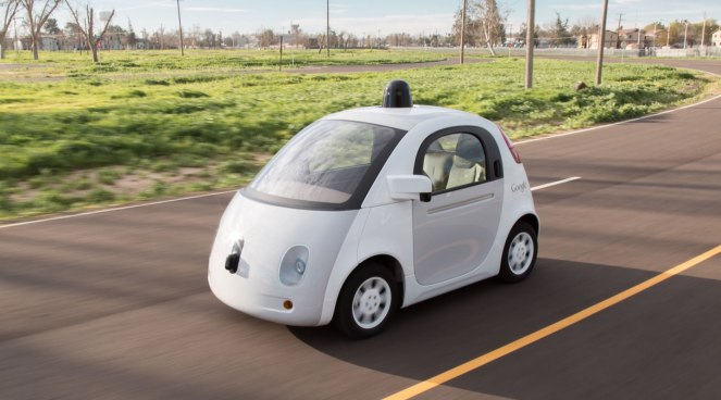
We have already heard about the future of releasing robots into our bloodstreams but we are also inching ever closer to releasing them onto our roads. In February 2012, Nevada became the first US state to allow the testing of autonomous cars on public roads.
Calm down though sci-fi fans, this doesn’t mean that whilst driving round Nevada you might pass a driver-less car, this just meant that they set out laws and guidelines that companies who want to test their self-drive vehicles must abide by. Future proofing, if you will. This law change started the ball rolling, with Florida, California and Michigan all following in Nevada’s footsteps. Or should I say tyre tracks? By the end of 2015 the UK, France and Switzerland have also brought out legislation on the testing of autonomous vehicles.
Even though things are moving in the right direction and there is much research being conducted in Universities, and by big companies such as Google, we are still a long way off from the technology being publicly available.
There is also some arguments over calling these vehicles autonomous, a word that implies the vehicle would be making decisions on its own and would be capable of doing so for long periods. Much of the technology that is currently being tested incorporates instructions from an information cloud and also from other vehicles close by. Many claim the term automated is a better fit.
Either way, it is possible that some time in the future you could be cruising round your home town and pass by a driver-less car, whether it will be ‘autonomous’ or ‘automated’ remains to be seen.
And finally …
Yay for the Pandas!

They are the face of wildlife conservation across the world and a brilliant source of entertainment on YouTube (Pandas playing on a slide is my personal favourite). In February 2015 the Chinese government in collaboration with WWF revealed that the wild Giant Panda population has increased by 17% in the last decade. A brilliant achievement in wild animal conservation. Lets hope the trend continues for the next decade.
Stay tuned for next months feature, looking back at some of the decades best scientific discoveries and achievements from the month of March.
Previous issues:
